 At Veritas Fidelitas, we practice fidelity to the truth by applying the most advanced neuroscience technologies in three principal areas:
Truth verification – establishes that you answered our battery of questions truthfully.
Recognition detection – indicates whether or not you recognize specific images (e.g. from a crime scene, which in turn indicates whether or not you were there).
Pain detection – identifies pain signatures in your brain to confirm that your pain is real, neither imagined nor feigned.
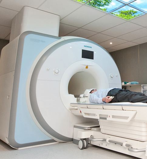
Our primary tool is the fMRI.
FUNCTIONAL MAGNETIC RESONANCE IMAGING: BASICS
fMRI is one of the most popular measurement techniques in cognitive neuroscience. It has been in use for about 20 years and is qualified as correlational because it records brain states in parallel with ongoing mental activity and/or behavior, thus permitting the establishment of correlational links between them. However, it does not allow researchers to establish a causal connection between brain states and behaviors or supposed mental processes. In most fMRI studies, brain states are the dependent variable measured during manipulation of the stimulus/task condition. Whether any specific local or systemic pattern of brain states is a necessary determinant of its associated behavior it cannot be determined with fMRI only. For this reason, fMRI is routinely used in basic research as a mainstay method to measure brain function and its data are often triangulated with data from complementary techniques (e.g., event-related potentials, transcranial magnetic stimulation), in a quest for converging evidence about mental processes and brain substrates.
As implied by its name, fMRI makes use of strong magnetic fields to create images of biological tissue. Depending on the pulse sequence of the electromagnetic fields it generates, an MRI scanner can detect different tissue properties and distinguish between tissue types. Scanners are used to acquire both brain structural information (e.g., allowing a fine distinction between white and gray matter, producing images of the static anatomy of the brain), and functional information such as measurements of local changes in blood oxygenation within the brain over time; the most common form of fMRI study. Because blood oxygenation levels change rapidly (i.e., after 1–2 s) following the activity of neurons in a brain region, fMRI allows researchers to localize brain activity on a second-by-second basis and within millimeters of its origin. These changes in blood oxygenation occur naturally and internally as part of normal brain physiology and, because the pulse sequence does not alter neuronal firing or blood flow, fMRI is considered a non-invasive technique.
Central to cognitive fMRI studies are the concepts of differences and similarities between maps of blood oxygenation level-dependent (BOLD) signal that are recorded in concomitance with different experimental conditions. In classical fMRI designs and in most of the available lie detection studies BOLD responses are evaluated in relative terms as the result of a contrast between two or more conditions. For example, maps of the BOLD signal that are recorded while a participant is lying can be contrasted with either maps recorded when the participant is at rest or when is telling the truth. Inferences about the neural correlates of lying are typically drawn from an analysis of the pattern of differences and/or similarities between BOLD signal maps across lying and not-lying conditions. In principle, any design difference (e.g., the use of a different stimulus or the requirement of additional mental operations given the same stimulus) between the lying condition and any other condition with which it is compared might lead to the recruitment of different brain regions to perform the task. Therefore, the more accurately the not-lying and lying conditions are matched, the more precise the conclusions that can be drawn about the neural correlates uniquely associated with lying. While this type of analysis is not the only possible or optimal way to draw informative inferences from fMRI data such contrast between conditions is a basic standard in fMRI research.
from Rusconi, E. (2013). Prospects of functional magnetic resonance imaging as lie detector. Frontiers in human neuroscience, 7, 594.doi:10.3389/fnhum.2013.00594
Below are videos on how two pioneering organizations use fMRI for lie detection.