
 A comprehensive collection of studies
on the neuroscience of truth
FULL REPORTS AVAILABLE UPON REQUEST
Truth Verification • Recognition Detection • Pain Detection
TRUTH VERIFICATION & LIE DETECTION
Basic and applied research on deception and its detection
Deception is a ubiquitous phenomenon in social interactions and has attracted a significant amount of research during the last decades. The majority of studies in this field focused on how deception modulates behavioral, autonomic, and brain responses and whether these changes can be used to validly identify lies. Especially the latter question, which historically gave rise to the development of psychophysiological “lie detection” techniques, has been driving research on deception and its detection until today. The detection of deception and concealed information in forensic examinations currently constitutes one of the most frequent applications of psychophysiological methods in the field. This Research Topic aims at bringing together contributions from researchers in experimental psychology, psychophysiology, and neuroscience focusing on the understanding of the broad concept of deception including the detection of concealed information, with respect to basic research questions as well as applied issues.
Deception is a complex social behavior which involves a set of higher cognitive functions. Studying this common phenomenon in humans has in all epochs been driven not merely by the wish to understand the underlying framework of cognitive functioning but rather by the ambition to detect deceptive behavior in criminal suspects. Thus, identifying valid indicators of deceptive behavior has always been in the focus of deception research. Such indicators can be defined in terms of specific behavior, physiological correlates, or content of verbal reports. The question of how validly each indicator allows for differentiating truthful and deceptive accounts is inherent in the majority of research efforts in this domain. Gamer, M., & Ambach, W. (2014). Deception research today. Frontiers in psychology, 5.
Functional MRI-based lie detection: scientific and societal challenges
Functional MRI (fMRI)-based lie detection has been marketed as a tool for enhancing personnel selection, strengthening national security and protecting personal reputations, and at least three US courts have been asked to admit the results of lie detection scans as evidence during trials. How well does fMRI-based lie detection perform, and how should the courts, and society more generally, respond? Here, we address various questions — some of which are based on a meta-analysis of published studies — concerning the scientific state of the art in fMRI-based lie detection and its legal status, and discuss broader ethical and societal implications. We close with three general policy recommendations. Farah, M. J., Hutchinson, J. B., Phelps, E. A., & Wagner, A. D. (2014). Functional MRI-based lie detection: scientific and societal challenges. Nature Reviews Neuroscience, 15(2), 123-131.
Cognitive control in belief-laden reasoning during conclusion processing: An ERP study
Belief bias is the tendency to accept conclusions that are compatible with existing beliefs more frequently than those that contradict beliefs. It is one of the most replicated behavioral findings in the reasoning literature. Recently, neuroimaging studies using functional magnetic resonance imaging (fMRI) and event-related potentials (ERPs) have provided a new perspective and have demonstrated neural correlates of belief bias that have been viewed as supportive of dual-process theories of belief bias. However, fMRI studies have tended to focus on conclusion processing, while ERPs studies have been concerned with the processing of premises. In the present research, the electrophysiological correlates of cognitive control were studied among 12 subjects using high-density ERPs. The analysis was focused on the conclusion presentation phase and was limited to normatively sanctioned responses to valid–believable and valid–unbelievable problems. Results showed that when participants gave normatively sanctioned responses to problems where belief and logic conflicted, a more positive ERP deflection was elicited than for normatively sanctioned responses to nonconflict problems. This was observed from -400 to -200 ms prior to the correct response being given. The positive component is argued to be analogous to the late positive component (LPC) involved in cognitive control processes. This is consistent with the inhibition of empirically anomalous information when conclusions are unbelievable. These data are important in elucidating the neural correlates of belief bias by providing evidence for electrophysiological correlates of conflict resolution during conclusion processing. Moreover, they are supportive of dual-process theories of belief bias that propose conflict detection and resolution processes as central to the explanation of belief bias. Luo, J., Liu, X., Stupple, E. N., Zhang, E., Xiao, X., Jia, L., & … Zhang, Q. (2013). Cognitive control in belief-laden reasoning during conclusion processing: An ERP study. International Journal Of Psychology, 48(3), 224-231. doi:10.1080/00207594.2012.677539
Scanning The Horizon: The Past, Present, and Future of Neuroimaging for Lie Detection In Court
The article focuses on the dangers and benefits of accurate lie detection and the constitutional and moral difficulties posed by lie detection technology. It explores the use of scientific methods and instruments for lie detection, particularly the active brain scan technology such as functional magnetic resonance imaging (fMRI). It also analyzes the admissibility of information derived from the said technology. Perhaps for the last time, academics have exclaimed to the world: We may finally have found a way to tell truth from lie! In a way that has not always been done in the past, the researchers working on this issue are expressing caution up-front. It is a hopeful sign. A breakthrough of the magnitude at issue demands great care and sober forethought. Nevertheless, revolutionary changes in the boundaries of human ability have always required both caution and the fortitude to accept and incorporate new paradigms. The arc of technological innovation falling rapidly into history behind us must be appreciated for what it tells us about our new trajectory. Now, more than ever before, humanity is called to adapt to the unfamiliar. If the promise of neuroimaging-based lie detection proceeds as seems likely, we will all be called to adapt to something wholly new and exciting—a world with verifiable truth. Brooks, S. J. (2012). Scanning The Horizon: The Past, Present, and Future of Neuroimaging for Lie Detection In Court. University Of Louisville Law Review, 51(2), 353-373.
The promises and perils of brain imaging technology: an ethical perspective
One area of incredible advances and promises within neurotechnology has been brain imaging technologies (BIT). But great promises often come along with great perils. In addition, given that BIT is not confined any more to the clinical setting the different ethical issues it can bring to the fore go beyond those that are regularly discussed in the medical context. For instance, who should see or have access to our brain image? Should neuroimages be used as evidence in court? Or for commercial issues? Here I will explore from a neuroethics perspective some of the promises and perils surrounding the uses of BIT outside the medical and research arenas. BITs are generally seen as technologies to monitor brain function. Among BIT one of the most portrayed in the media and public discourse is functional Magnetic Resonance Imaging (fMRI), but there are other imaging technologies, such as Positron emission Tomography (PET) and electroencephalography EEG. BIT has unique capabilities inasmuch as it can help us to understand the brain in detail and depth that previous technologies and methods have not enabled us to achieve before. It can also help us to understand how the brain develops, changes and wires itself, which in turn can shed light into our understanding of human behavior and cognition. So among the promises of BIT we found claims related to it helping us to better monitor and diagnose neuro-related diseases and conditions, which could have a positive impact for society. However, we should bear in mind that at present BIT is far from being perfect, and its reliability is often questioned, being charged with issues related to overinference, neuroasthetics, confirmation bias, and trait state confusion (Farah, 2010, Wolpe et al, 2005). There is another source of concern, namely the way neuroimaging results are interpreted. Some people have seen the potential of BITs to tell us something about mental states which can then be used for: neuromarketing, lie detection (brain reading), and assessment of potential dangerousness. Others have seen it as the analogous to genotyping, in which through our brainotype we will be able to measures, for instance how personality traits are reflected in the brain’s functional architecture and as such determine the kind of persons we are or how different brain areas at work can tell us about morally relevant intuitions and those that are not (Levy, 2011). However, it is questionable the ability of brain imaging to provide an objective interpretation of complex social behavior (Phelps and Thomas, 2003), such as whether someone is lying (Wolpe and Foster, 2005), likely to show dangerous behavior or about our morality (Cabrera, 2011). It is one thing to use BIT to predict the likelihood of someone developing a certain neurological condition and another thing to say that we can use BIT to predict dangerousness. Imaging technologies can tell us that certain mental activity correlates with activity in certain brain region, but not whether the activity is actually the result the mental activity itself. Now, even if we ever developed a BIT with which we feel comfortable to use for assessing whether someone has committed or not a crime, or whether that someone shows a ‘dangerous brain’ and as such it is unsuitable for release, it will not make much sense to use it for such purposes unless we already have a program in place to deal with the positive cases we might encounter. We have to remain cautious in the ways we use neuroimaging technologies but also in the way we interpret neuroimages. The new understandings that BIT promise to give us can certainly help us to capture the dynamic and complex ways of our mental life, including our intuitions, beliefs, desires as they tell us about their neural/psychological bases and the way they are influenced by reasoning, emotion, and social influences; but these new understandings could also reduce complex human behaviors to just certain brain activities while stigmatizing other important resources and elements of our mental processes (Cabrera, 2011). As Martha Farah (2010) has put it “some of the most profound ethical challenges from neuroscience come not from new technologies but from new understandings”. Thus, if we want to ensure that these technologies are used for the benefit of our society, then we have to start questioning ourselves about the practical value of neuroscientific knowledge and how we should best realize it. Cabrera, L. Y. (2013). The promises and perils of brain imaging technology: an ethical perspective. Proceedings Of The Physiological Society, 185P-186P.
Weighing the admissibility of fMRI technology under FRE 403
The article describes the anatomy of a lie and highlights several technologies used to detect lie including functional magnetic resonance imaging (fMRI), polygraph and control question test (CQT). It discusses the evidentiary rules governing the admissibility of these technologies and analyzes whether admissibility of fMRI deception detection technology fulfills the governing standards of scientific expert testimony. Amirian, J. (2013). Weighing the admissibility of fMRI technology under FRE 403. Fordham Urban Law Journal, 41(2), 715-770.
Lies, damned lies, and physiology
The use of biological evidence in Court is now commonplace. While traditional forensic evidence remains extremely valuable, the holy grail of a foolproof lie detector has so far eluded law enforcement agencies. To this end, research efforts continue with a view to identifying a biological signature of deception. Mohammed and colleagues (Mohammed et al. 2006) have suggested that in a lie, the sequence of events is as follows. The question is put to the subject. The subject processes the information, recognises the truthful answer, suppresses the truthful answer, considers an appropriate untrue answer and then articulates the appropriate untrue answer. The mental effort needed in truth telling is seen to be less – as the subject simply has to understand the question, recall the truthful answer and articulate it. Mohammed and co- workers (Mohammed et al. 2006) have further opined that fourteen areas of the brain “light up” in deception whereas only seven are involved in telling the truth. fMRI identifies the BOLD response to the additional cognitive effort involved in deception. Green, M. (2011). Lies, damned lies and physiology. Biologist, 58(1), 29-32.
Prospects of functional magnetic resonance imaging as lie detector
Following the demise of the polygraph, supporters of assisted scientific lie detection tools have enthusiastically appropriated neuroimaging technologies “as the savior of scientifically verifiable lie detection in the courtroom” (Gerard, 2008: 5). These proponents believe the future impact of neuroscience “will be inevitable, dramatic, and will fundamentally alter the way the law does business” (Erickson, 2010: 29); however, such enthusiasm may prove premature. For in nearly every article published by independent researchers in peer reviewed journals, the respective authors acknowledge that fMRI research, processes, and technology are insufficiently developed and understood for gatekeepers to even consider introducing these neuroimaging measures into criminal courts as they stand today for the purpose of determining the veracity of statements made. Regardless of how favorable their analyses of fMRI or its future potential, they all acknowledge the presence of issues yet to be resolved. Even assuming a future where these issues are resolved and an appropriate fMRI lie-detection process is developed, its integration into criminal trials is not assured for the very success of such a future system may necessitate its exclusion from courtrooms on the basis of existing legal and ethical prohibitions. In this piece, aimed for a multidisciplinary readership, we seek to highlight and bring together the multitude of hurdles which would need to be successfully overcome before fMRI can (if ever) be a viable applied lie detection system. We argue that the current status of fMRI studies on lie detection meets neither basic legal nor scientific standards. We identify four general classes of hurdles (scientific, legal and ethical, operational, and social) and provide an overview on the stages and operations involved in fMRI studies, as well as the difficulties of translating these laboratory protocols into a practical criminal justice environment. It is our overall conclusion that fMRI is unlikely to constitute a viable lie detector for criminal courts. Rusconi, E. (2013). Prospects of functional magnetic resonance imaging as lie detector. Frontiers in human neuroscience, 7, 594.doi:10.3389/fnhum.2013.00594
Detecting Deception Using Functional Magnetic Resonance Imaging
The ability to accurately detect deception is presently very limited. Detecting deception might be more accurately achieved by measuring the brain correlates of lying in an individual. In addition, a method to investigate the neurocircuitry of deception might provide a unique opportunity to test the neurocircuitry of persons in whom deception is a prominent component (i.e., conduct disorder, antisocial personality disorder, etc.). In this study, we used functional magnetic resonance imaging (fMRI) to show that specific regions were reproducibly activated when subjects deceived. Subjects participated in a mock crime stealing either a ring or a watch. While undergoing an fMRI, the subjects denied taking either object, thus telling the truth with some responses, and lying with others. A Model-Building Group (MBG, n = 30) was used to develop the analysis methods, and the methods were subsequently applied to an independent Model-Testing Group (MTG, n = 31). We were able to correctly differentiate truthful from deceptive responses, correctly identifying the object stolen, for 93% of the subjects in the MBG and 90% of the subjects in the MTG. This is the first study to use fMRI to detect deception at the individual level. Further work is required to determine how well this technology will work in different settings and populations. Kozel, F. A., Johnson, K. A., Mu, Q., Grenesko, E. L., Laken, S. J., & George, M. S. (2005). Detecting deception using functional magnetic resonance imaging. Biological psychiatry, 58(8), 605-613.
Telling truth from lie in individual subjects with fast event-related fMRI
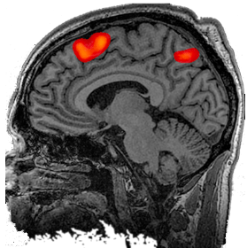
Deception is a clinically important behavior with poorly understood neurobiological correlates. Published functional MRI (fMRI) data on the brain activity during deception indicates that, on a multisubject group level, lie is distinguished from truth by increased prefrontal and parietal activity. These findings are theoretically important; however, their applied value will be determined by the accuracy of the discrimination between single deceptive and truthful responses in individual subjects. This study presents the first quantitative estimate of the accuracy of fMRI in conjunction with a formal forced-choice paradigm in detecting deception in individual subjects. We used a paradigm balancing the salience of the target cues to elicit deceptive and truthful responses and determined the accuracy of this model in the classification of single lie and truth events. The relative salience of the task cues affected the net activation associated with lie in the superior medial and inferolateral prefrontal cortices. Lie was discriminated from truth on a single-event level with an accuracy of 78%, while the predictive ability expressed as the area under the curve (AUC) of the receiver operator characteristic curve (ROC) was 85%. Our findings confirm that fMRI, in conjunction with a carefully controlled query procedure, could be used to detect deception in individual subjects. Salience of the task cues is a potential confounding factor in the fMRI pattern attributed to deception in forced choice deception paradigms. Langleben, D. D., Loughead, J. W., Bilker, W. B., Ruparel, K., Childress, A. R., Busch, S. I., & Gur, R. C. (2005). Telling truth from lie in individual subjects with fast event‐related fMRI. Human brain mapping, 26(4), 262-272.
Current research and potential applications of the concealed information test: an overview
Research interest in psychophysiological detection of deception has significantly increased since the September 11 terror attack in the USA. In particular, the concealed information test (CIT), designed to detect memory traces that can connect suspects to a certain crime, has been extensively studied. In this paper I will briefly review several psychophysiological detection paradigms that have been studied, with a focus on the CIT. The theoretical back- ground of the CIT, its strength and weaknesses, its potential applications as well as research finings related to its validity (based on a recent meta-analytic study), will be discussed. Several novel research directions, with a focus on factors that may affect CIT detection in realistic settings (e.g., memory for crime details; the effect of emotional stress during crime execution) will be described. Additionally, research focusing on mal-intentions and attempts to detect terror networks using information gathered from groups of suspects using both the standard CIT and the searching CIT will be reviewed. Finally, implications of current research to the actual application of the CIT will be discussed and several recommendations that can enhance the use of the CIT will be made. Ben-Shakhar, G. (2012). Current research and potential applications of the concealed information test: an overview. Frontiers in psychology, 3.
The anterior cingulate cortex and pain processing
The neural network that contributes to the suffering which accompanies persistent pain states involves a number of brain regions. Of primary interest is the contribution of the cingulate cortex in processing the affective component of pain. The purpose of this review is to summarize recent data obtained using novel behavioral paradigms in animals based on measuring escape and/or avoidance of a noxious stimulus. These paradigms have successfully been used to study the nature of the neuroanatomical and neurochemical contributions of the anterior cingulate cortex (ACC) to higher order pain processing in rodents. Fuchs, Perry N., et al. “The anterior cingulate cortex and pain processing.” Frontiers in integrative neuroscience 8 (2014).
Methodological aspects of connectivity analyses of the neuromatrix of pain
Pain is a subjective phenomenon, produced by the interactions of a complex and dynamic network of brain structures termed the brain’s neuromatrix of pain (BNMP), and composed of different cortical and subcortical brain areas. Although EEG/MEG and fMRI allow measurement and localization of brain activity on different spatial and temporal scales, interactions between activated brain areas cannot be directly measured. Their study requires sophisticated analysis tools and modelling that use measured brain activity as input data. Witte, H. Methodological aspects of connectivity analyses of the neuromatrix of pain. Fechner Day 2013, 18.

Beyond Patient Reported Pain: Perfusion Magnetic Resonance Imaging Demonstrates Reproducible Cerebral Representation of Ongoing Post-Surgical Pain
Development of treatments for acute and chronic pain conditions remains a challenge, with an unmet need for improved sensitivity and reproducibility in measuring pain in patients. Here we used pulsed-continuous arterial spin-labelling [pCASL], a relatively novel perfusion magnetic-resonance imaging technique, in conjunction with a commonly-used post-surgical model, to measure changes in regional cerebral blood flow [rCBF] associated with the experience of being in ongoing pain. We demonstrate repeatable, reproducible assessment of ongoing pain that is independent of patient self-report. In a cross-over trial design, 16 participants requiring bilateral removal of lower-jaw third molars underwent pain-free pre-surgical pCASL scans. Following extraction of either left or right tooth, repeat scans were acquired during post-operative ongoing pain. When pain-free following surgical recovery, the pre/post-surgical scanning procedure was repeated for the remaining tooth. Voxelwise statistical comparison of pre and post-surgical scans was performed to reveal rCBF changes representing ongoing pain. In addition, rCBF values in predefined pain and control brain regions were obtained. rCBF increases (5–10%) representing post-surgical ongoing pain were identified bilaterally in a network including primary and secondary somatosensory, insula and cingulate cortices, thalamus, amygdala, hippocampus, midbrain and brainstem (including trigeminal ganglion and principal-sensory nucleus), but not in a control region in visual cortex. rCBF changes were reproducible, with no rCBF differences identified across scans within-session or between post-surgical pain sessions. This is the first report of the cerebral representation of ongoing post-surgical pain without the need for exogenous tracers. Regions of rCBF increases are plausibly associated with pain and the technique is reproducible, providing an attractive proposition for testing interventions for on-going pain that do not rely solely on patient self-report. Our findings have the potential to improve our understanding of the cerebral representation of persistent painful conditions, leading to improved identification of specific patient sub-types and implementation of mechanism-based treatments. Howard, M. A., Krause, K., Khawaja, N., Massat, N., Zelaya, F., Schumann, G., … & Renton, T. F. (2011). Beyond patient reported pain: perfusion magnetic resonance imaging demonstrates reproducible cerebral representation of ongoing post-surgical pain. PloS one, 6(2), e17096.